

Elbow
Anatomy
The elbow is a complex joint formed by the articulation of three bones –the humerus, radius and ulna. The elbow joint helps in bending or straightening of the arm to 180 degrees and assists in lifting or moving objects.
The bones of the elbow are supported by
- Ligaments and tendons
- Muscles
- Nerves
- Blood vessels
Bones and Joints of the elbow joint:
The elbow joint is formed at the junction of three bones:
- The Humerus (upper arm bone) forms the upper portion of the joint. The lower end of the humerus divides in to two bony protrusions known as the medial and lateral epicondyles which can be felt on either side of the elbow joint.
- The Ulna is the larger bone of the forearm located on the inner surface of the joint. The curved shape of the ulna articulates with the humerus.
- The Radius is the smaller bone of the forearm situated on the outer surface of the joint. The head of the radius is circular and hollow which allows movement with the humerus. The connection between the ulna and radius helps the forearm to rotate.
The elbow consists of three joints from articulation of the three bones namely:
- Humero-ulnar joint is formed between the humerus and ulna and allows flexion and extension of the arm.
- Humero-radial joint is formed between the radius and humerus, and allows movements like flexion, extension, supination and pronation.
- Radio-ulnar joint is formed between ulna and radius bones, and allows rotation of the lower arm.
Articular cartilage lines the articulating regions of the humerus, radius and ulna. It is a thin, tough, flexible, and slippery surface that acts as a shock absorber and cushion to reduce friction between the bones. The cartilage is lubricated by synovial fluid, which further enables the smooth movement of the bones.
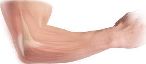
Muscles of the Elbow Joint
There are several muscles extending across the elbow joint that help in various movements. These include the following:
- Biceps brachii: upper arm muscle enabling flexion of the arm
- Triceps brachii: muscle in the back of the upper arm that extends the arm and fixes the elbow during fine movements
- Brachialis: upper arm muscle beneath the biceps which flexes the elbow towards the body
- Brachioradialis: forearm muscle that flexes, straightens and pulls the arm at the elbow
- Pronator teres: this muscle extends from the humeral head, across the elbow, and towards the ulna, and helps to turn the palm facing backward
- Extensor carpi radialis brevis: forearm muscle that helps in movement of the hand
- Extensor digitorum: forearm muscle that helps in movement of the fingers
Elbow joint ligaments and tendons:
The elbow joint is supported by ligaments and tendons, which provide stability to the joint.
Ligaments are a group of firm tissues that connect bones to other bones. The most important ligaments of the elbow joint are the:
- Medial or ulnar collateral ligament: comprised of triangular bands of tissue on the inner side of the elbow joint.
- Lateral or radial collateral ligament: a thin band of tissue on the outer side of the elbow joint.
Together, the medial and lateral ligaments are the main source of stability and hold the humerus and ulna tightly in place during movement of the arm.
- Annular ligament: These are a group of fibers that surrounds the radial head, and holds the ulna and radius tightly in place during movement of the arm.
The ligaments around a joint combine to form a joint capsule that contains synovial fluid.
Any injury to these ligaments can lead to instability of the elbow joint.
Tendons are bands of connective tissue fibers that connect muscle to bone. The various tendons which surround the elbow joint include:
- Biceps tendon: attaches the biceps muscle to the radius, allowing the elbow to bend
- Triceps tendon: attaches the triceps muscle to the ulna, allowing the elbow to straighten
Nerves of the elbow joint:
The main nerves of the elbow joint are the ulnar, radial and median nerves. These nerves transfer signals from the brain to the muscles that aid in elbow movements. They also carry the sensory signals like touch, pain, and temperature back to the brain.
Any injury or damage to these nerves causes pain, weakness or joint instability.
Blood vessels:
Arteries are blood vessels that carry oxygen-pure blood from the heart to the hand. The main artery of the elbow is the brachial artery that travels across the inside of the elbow and divides into two small branches below the elbow to form the ulnar and the radial artery.
Elbow Tendon and Ligament Repair
The elbow is a complex joint of the upper limb formed by the articulation of the long bone of the upper arm or humerus and the two bones of the forearm, namely, radius and ulna. It is one of the important joints of the upper limb and is involved in basic movements such as flexion and extension of the upper limb and rotation of the forearm.
The elbow joint is supported by the ulnar collateral ligament, radial collateral ligament and the annular ligament. These ligaments provide stability and strength to the elbow joint.
The elbow joint also has the attachment of the common flexor and common extensor tendons. These groups of muscle assist in rotational movement of the forearm as well as the movements of the wrist and hand.
Injuries
The common conditions affecting the tendons around the elbow joint include tennis elbow and golfer’s elbow which result from an overuse injury to the tendons or result from repetitive activities such as sports, mechanical activities or weight lifting.
The ligaments around the elbow may be injured secondary to a sprain, rupture, trauma or any accident. The sprain or trauma may result from repetitive stress, overuse or a direct injury.
Symptoms
The common symptoms of injury to the elbow joint and its surrounding structures include swelling and pain, which may extend from the elbow to the forearm and palm and be aggravated by movements of the wrist. Sometimes instability of the joint may also be seen.
Surgical procedure
Tendon repair
The repair of the damaged tendon is broadly classified into two types- tendon debridement and tendon release.
Tendon debridement
This procedure is commonly used in for the management of tendinitis. In this procedure, the surgeon removes any damaged tissue from the tendon and cleans the tendon.
Tendon release
It is the most commonly used surgery for tendon repair. In this procedure, the surgeon locates the attachment of the extensor or flexor tendon on the elbow and splits the damaged tendon as well as removes the scar tissue or other overgrowth, around the tendon. Sometimes the loose end of the tendon may be sutured to the surrounding connective tissue (fascia).
Ligament reconstruction
Ligament reconstruction is considered in patients with ligament rupture. Your surgeon will make an incision over the elbow. Care is taken to move muscles, tendons and nerves out of the way. The donor tendon is harvested from either the forearm or below the knee. Your surgeon drills holes into the bones of the upper arm and the forearm, around the elbow joint. The donor tendon is inserted through the drilled holes in a pattern like that of the original ligament complex. The tendon is then attached to the bone surfaces with special sutures. The incision is closed with sutures and covered with sterile dressings. A splint is applied to support the elbow for a few weeks. After the surgery, you might be advised for regular follow-up and for a rehabilitation program for a better and quicker recovery.
Complications
Common complications of the elbow ligament and tendon repair surgeries include infection, injury to the adjacent nerve and blood vessels, and a loss of strength or flexibility of the elbow joint.
Rehabilitation
The success of the surgery depends on the post-operative rehabilitation program which includes the use of a removable splint immediately after surgery as well as ice therapy, electrical stimulation and massage for reducing pain, swelling or muscle spasm. Isometric exercises, strengthening and range of motion exercises may be useful for long term rehabilitation.